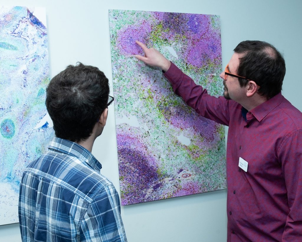
The sunlit atrium at La Jolla Institute for Immunology (LJI) is now a place to bask in the beauty of science. The Institute recently unveiled a new “pocket” art gallery to showcase breathtaking images of immune cells at work.
The images—captured through high-powered microscopes—reveal a secret, silent world. B cells flock to indigo-stained follicles to hone their virus-fighting skills. A rainbow of immune cells, epithelial cells, and neurons come together in the jagged terrain of the small intestine.
“The Immune Landscape” art gallery was made possible thanks to the generous support of LJI Board Director Barbara Donnell, M.A., a long-time advocate for scientific advancement and career development programs at LJI. Donnell says that touring LJI’s Microscopy and Histology Core Facility inspired her to look for ways to highlight the intersection between art and science.
“I love that we can zoom into cells like this,” says Donnell. “A picture is worth a thousand words, and to get those images on the walls really brings science to life.”
Zbigniew Mikulski, Ph.D., LJI Microscopy Core Director, led the effort to select and prepare images for the gallery. His team works with an advanced multiplex imaging technology, which enables them to partner with LJI labs to study the immune system’s response to cancers, autoimmune disease, and infectious disease. This cutting-edge technology was acquired by LJI thanks to a generous gift from Michael and Ellise Coit.
Mikulski is eager for non-scientists to explore the gallery and see what cutting-edge imaging reveals about the body. “We want people to see that we’re beautiful not just on the outside but on the inside too,” says Mikulski. “There is structure and harmony in our tissues. Every cell in the body evolved to have a specific role. It’s a system. It all works together.”
The gallery has also proven eye-opening for scientists who don’t normally see tissue samples at such a huge scale. “Most of them are like, ‘Whoa! What’s that?'” says Mikulski. “These images are rich with information, and they do a good job of showing the landscape of the immune system.”
Donnell hopes the new art gallery will also help LJI scientists feel celebrated and motivated to work toward new discoveries. She’s currently working with LJI staff to produce a video to highlight LJI’s microscopy work and snapshots views of the immune system in action.
“I think it means something for scientists to see their work displayed like this,” says Donnell. “I hope the art gallery leads to new appreciation for this work—and awe, wonder, and inspiration.”
Learn more about LJI’s efforts to showcase art and science:
All about LJI’s Student Art Competition
Meet an artist behind eye-catching scientific journal covers